CIPTA®2 – The unique cloud software for custom corneal surgery


CIPTA®2 is the ultimate cloud software conceived to:

  • plan and overview customized surgical treatments anywhere at anytime on the internet;
  • have remote access to any device of the 4D Suite™;
  • allow simultaneous interaction between multiple Users;
  • have remote access to iVerify® and iTicket web-app;
  • be mobile friendly.

CIPTA®2 supports customized surgical plan for:

  • Monofocal and Multifocal refractive treatments for Not Irregular Disorders (NID) as Myopia, Hyperopia, Astigmatism, Myopic Astigmatism, Hyperopic Astigmatism and Mixed Astigmatism;
  • Therapeutic treatments for Stable Irregular Disorders (SID) as Irregular Astigmatism, Decentrations, Scars and Corneal Opacities;
  • Therapeutic treatments for Ectatic Irregular Disorders (EID) as Keratoconus and Pellucida, combined with customized or standard crosslinking treatments to stabilize the corneal structure.

Key Features

icon raytracing 64x64

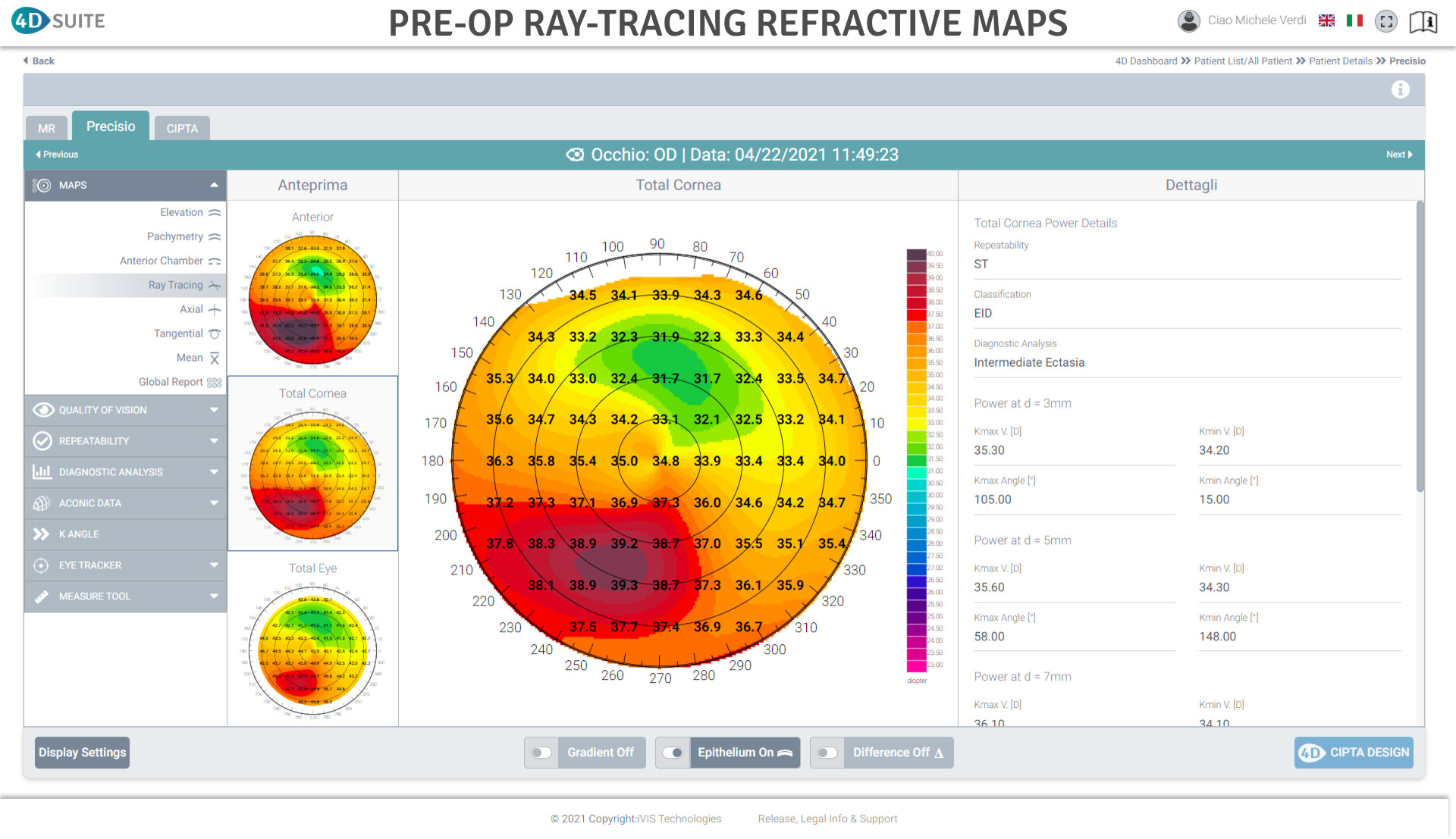
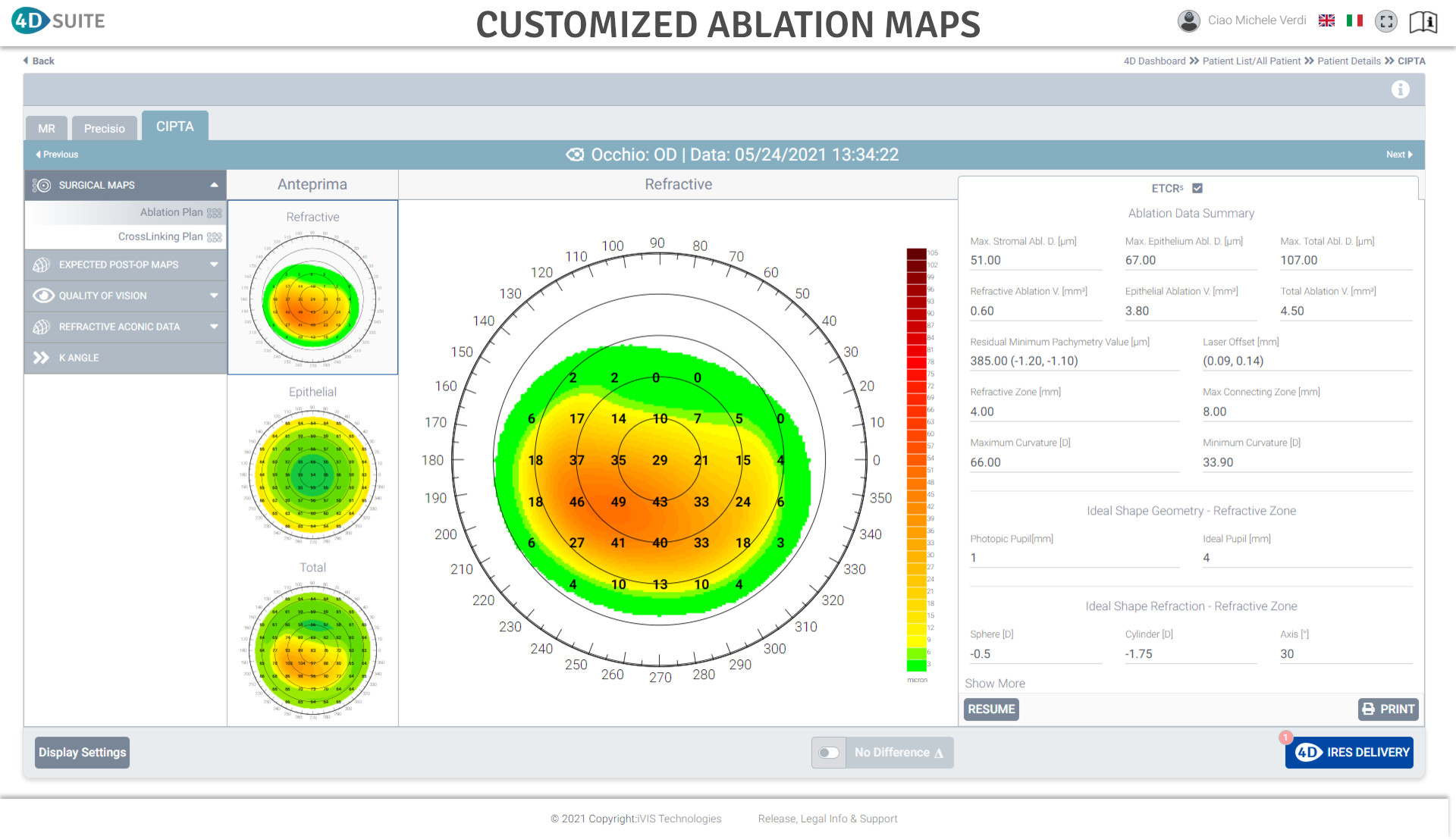
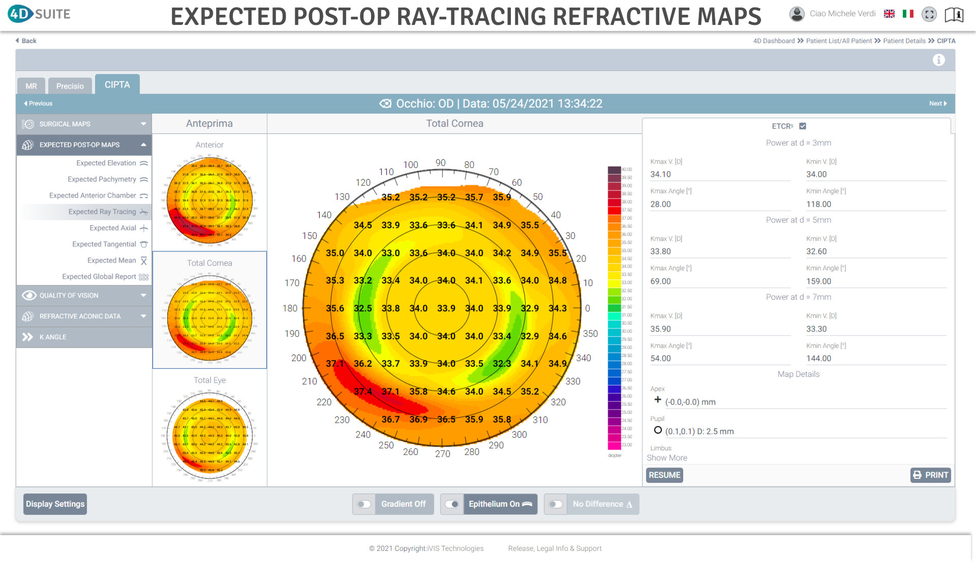
Ray Tracing customization of the ablation profile

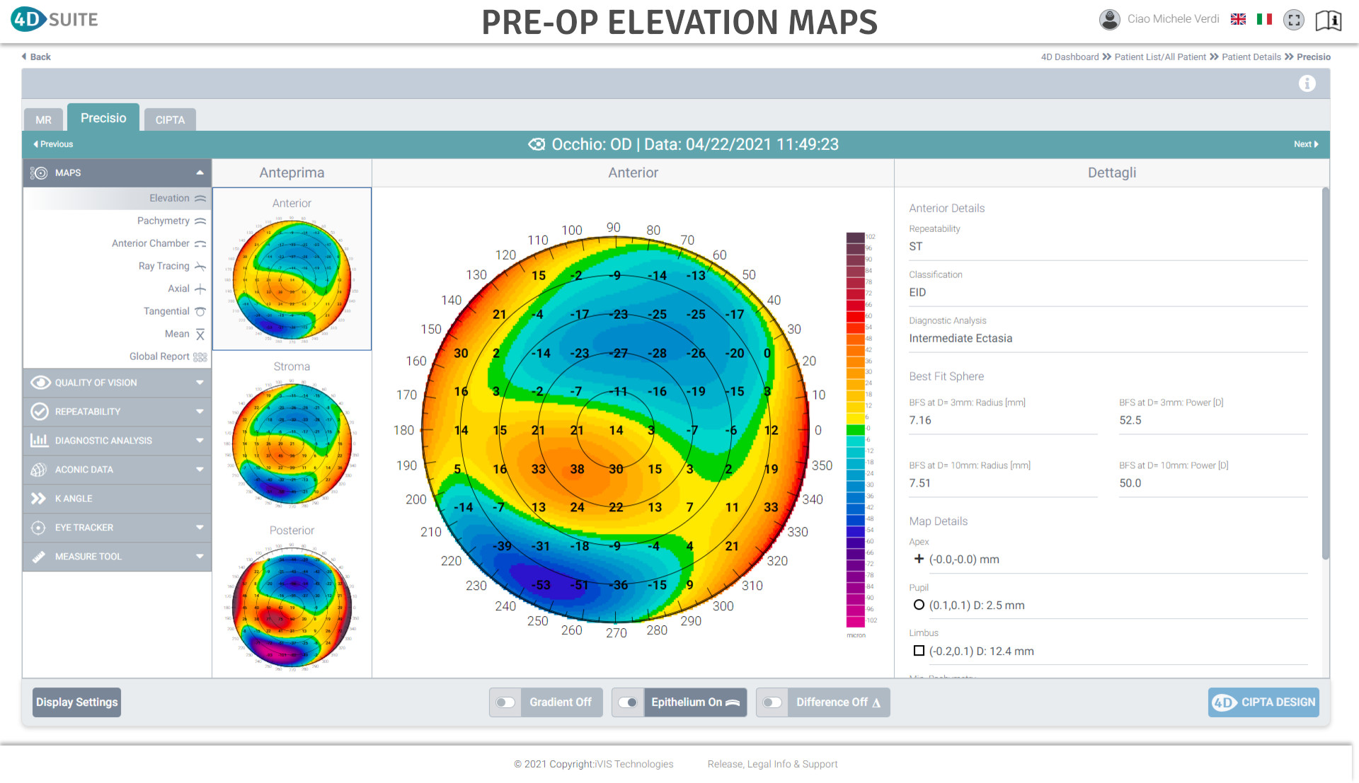
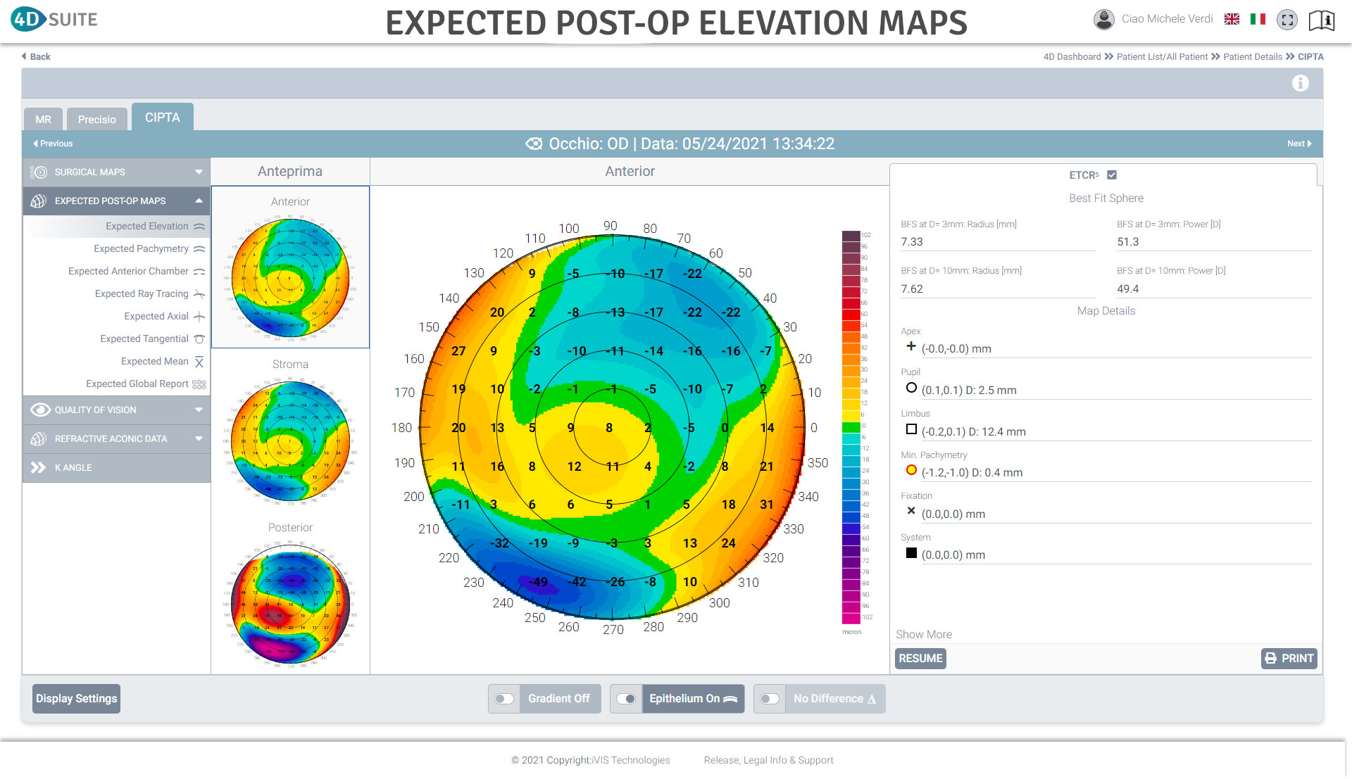
Refractive contribution of the posterior corneal surface

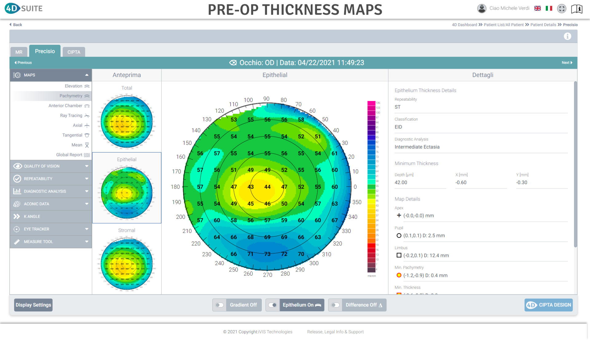
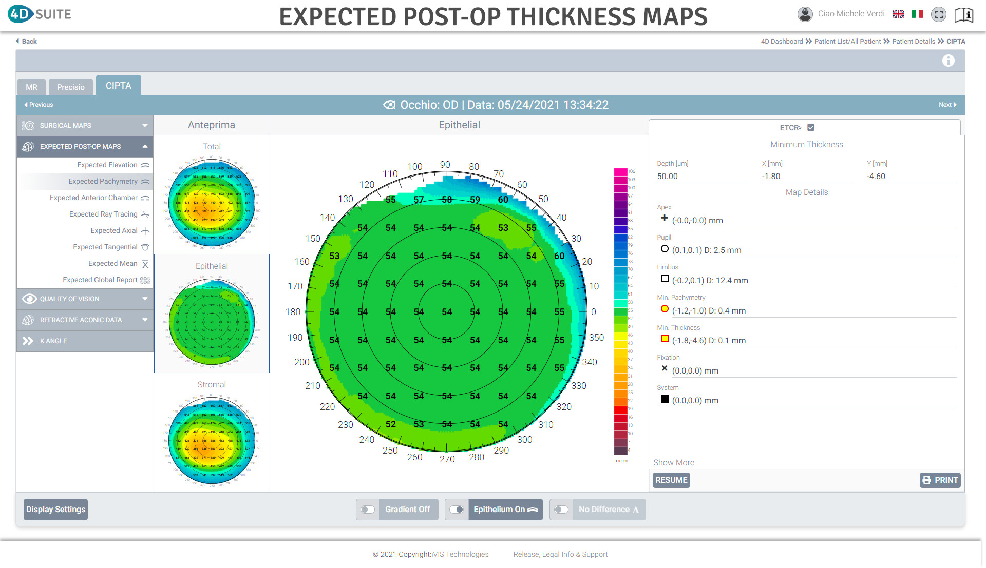
Customized epithelium and stromal ablation plan

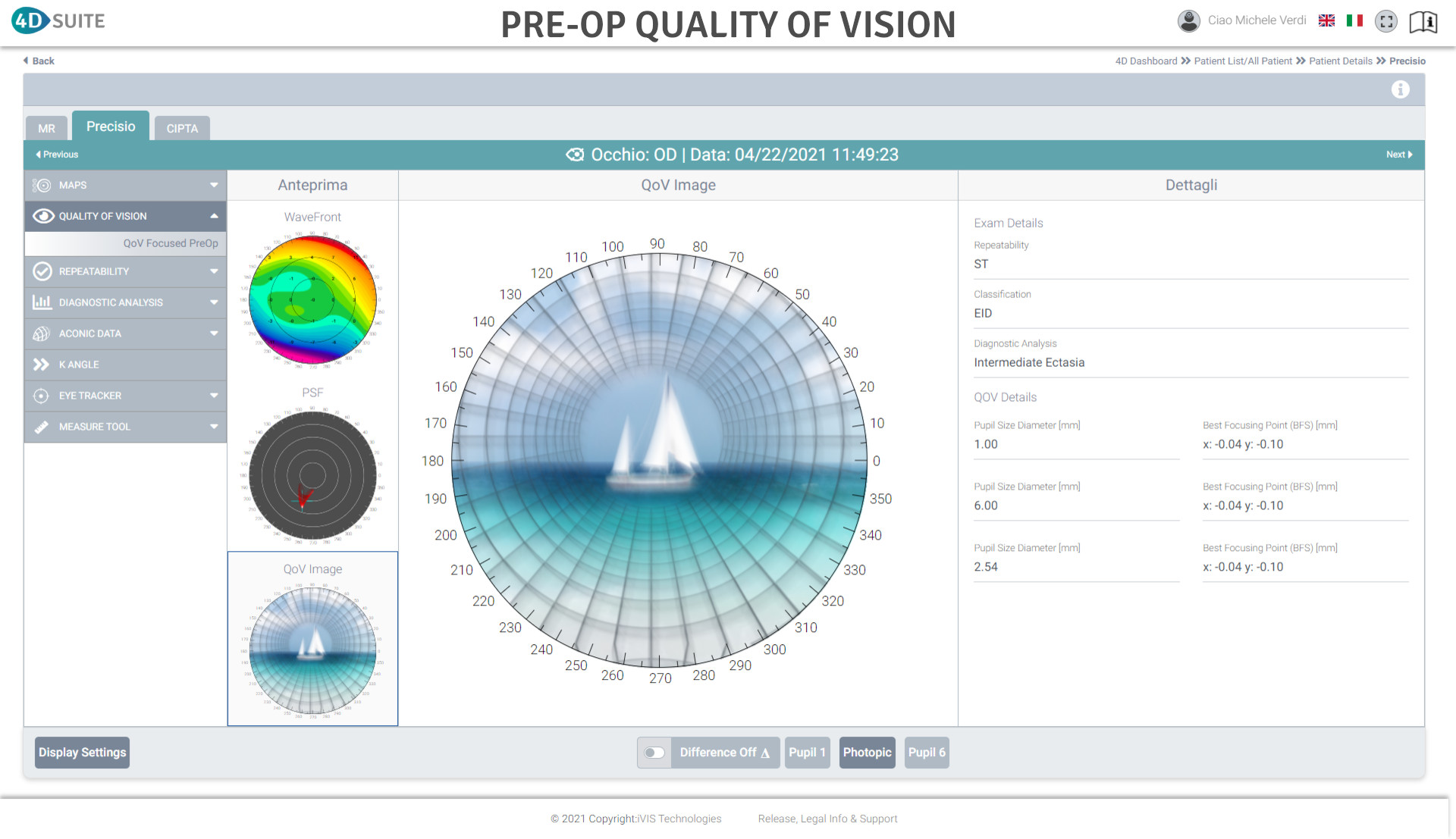
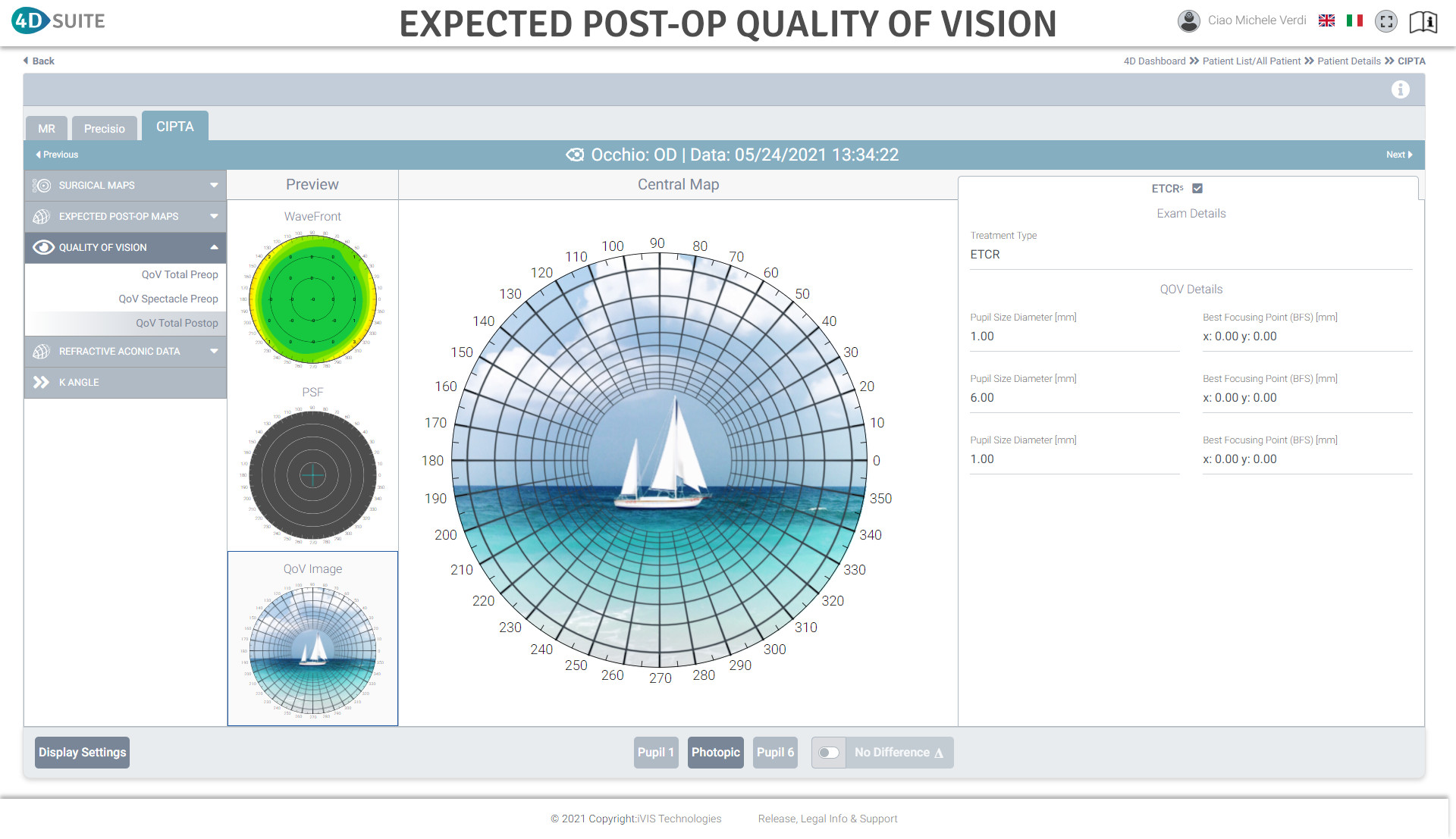
Quality of Vision analysis

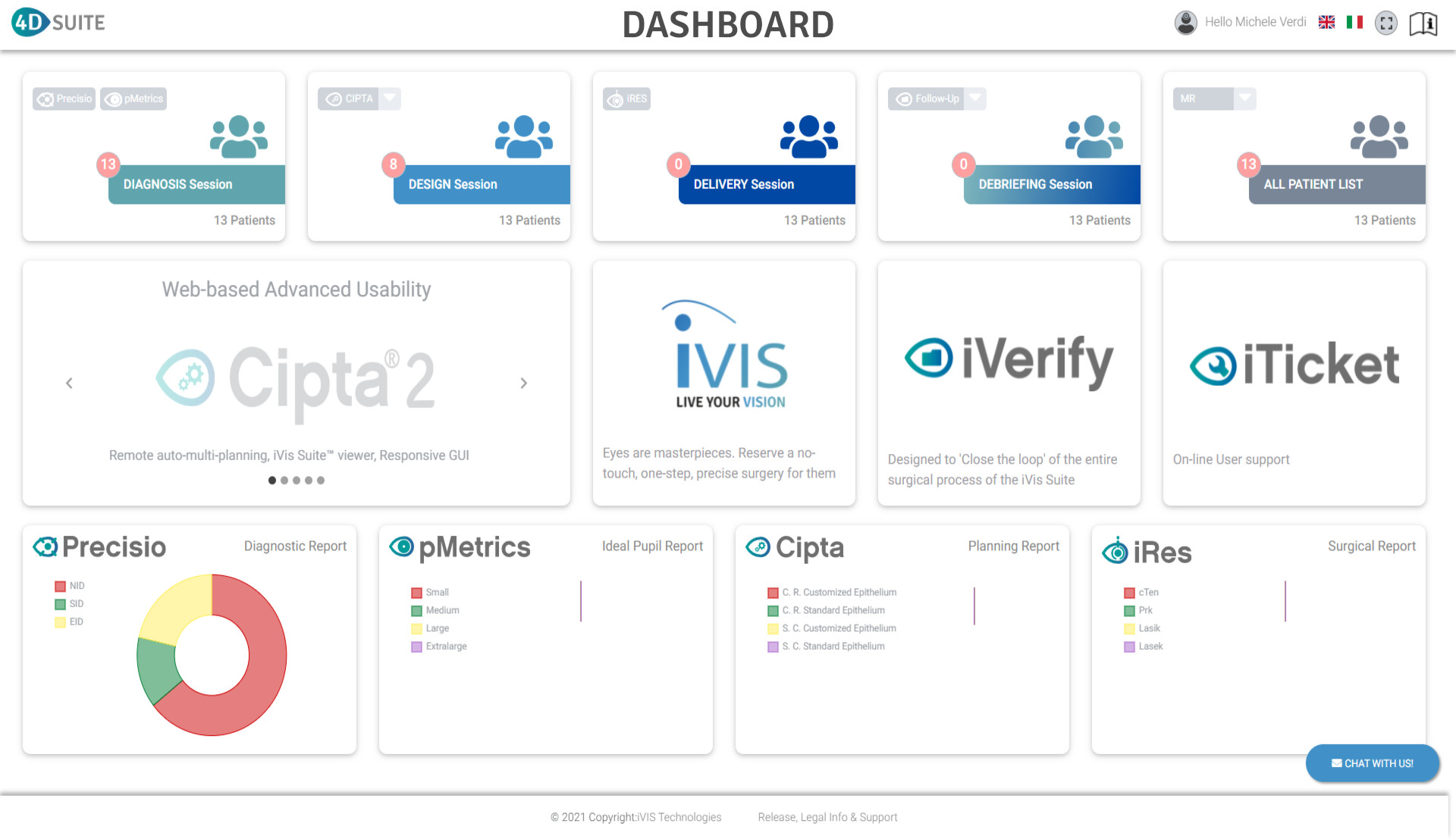
Remote Access to the 4D Suite™

real time

Real time upgrades, updates and data sharing

Fully automated surgical planning

User friendly Interface

Principles

The tissue to be removed is defined by the intersection between the real cornea shape detected by Precisio® and the Ideal shape tailored by a patented Ray Tracing process

The refractive zone is limited by the projection of the Ideal Pupil, defined by pMetrics®, onto the corneal shape

The connecting zone between the refractive zone and the untouched cornea is customized to grant a Constant slope

Clinical Application

Customized corneal ablation for the treatment of refractive disorders

Customized corneal ablation for the treatment of corneal stable pathologies

Customized corneal ablation and crosslinking for the treatment of ectatic corneal pathologies

Customized first order correction for strabismus

Customized first order correction for macula degeneration

Exam Outputs

Stromal ablation map

Epithelial ablation map

Total ablation map

Crosslinking treatment map

Expected post-op Elevation maps: anterior, stromal and posterior

Expected post-op Thicnkess map: epithelium, stromal and total

Expected post-op Ray Tracing refractive map: anterior, stromal, posterior and total

Expected post-op Axial refractive map: anterior, stromal, posterior and total

Expected post-op Tangential refractive map: anterior, stromal, posterior and total

Expected post-op Mean power refractive map: anterior, stromal, posterior and total

Expected post-op Wavefront map

Expected post-op PSF map

Expected post-op Quality of Vision images