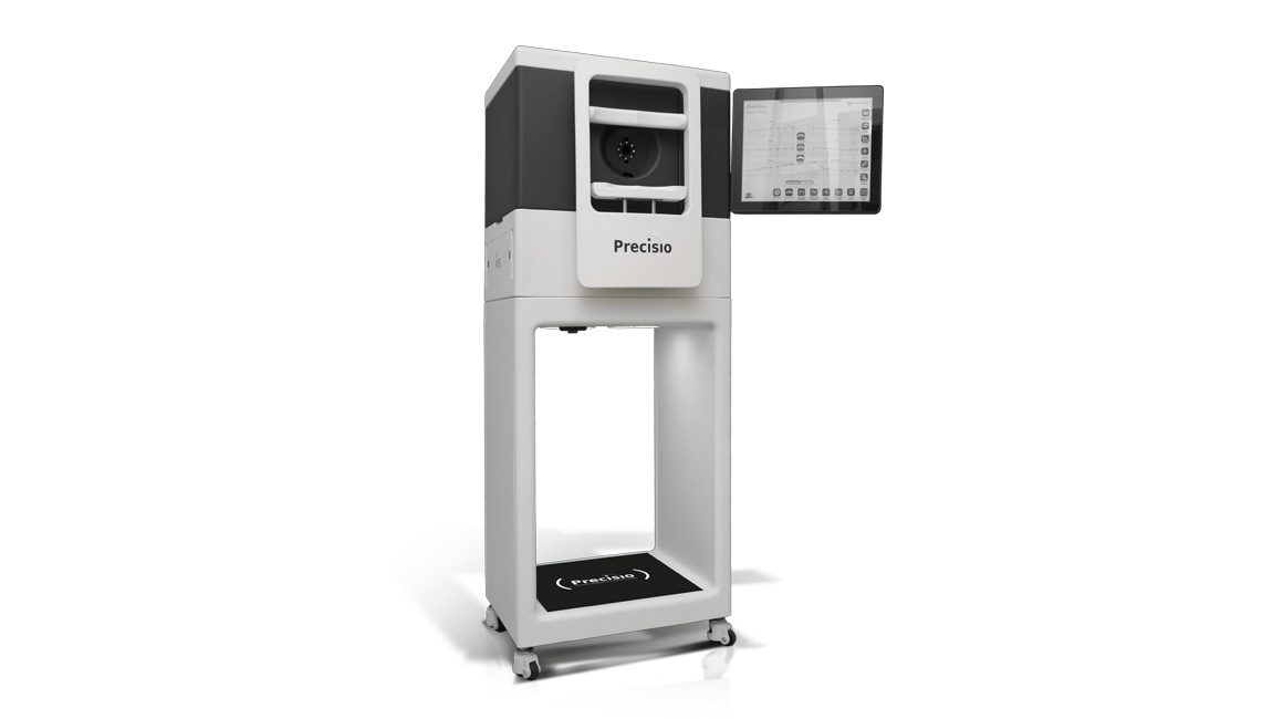
Precisio® – High Resolution Tomographer
The unique Tomographer for fully customized eye surgery and highly detailed diagnosis.
Designed in-house and fully integrated into the iVis Suite™, Precisio®2 is iVis’ second generation high resolution Corneal Tomographer. It delivers true HD morphological and refractive maps and ID data for eye registration with a great repeatability of a few microns.
Precisio®2 incorporates a patented ultra-thin blue scanning laser slit, to allow a lacrimal film-independent detection of fully registered and validated epithelial maps up to 10.0 mm of corneal diameter.
Precisio®2 is linked to the web-based iVerify® application, to grant an automated and objective debriefing process, to allow clinical follow-up of corneal pathologies, to analyse morphological and refractive corneal surgical outcomes, and to allow surgical follow-up of the customized refractive and therapeutic treatments.
Precisio®2 incorporates a 6D eye tracking sub-system to allow an accurate registration of 60 HD cross-section images acquired in less than 1 sec, thus providing morphological and refractive data of each and every corneal sublayer, including the iris plane, with unmatched accuracy and repeatability.
Thanks to a proprietary Ray-Tracing technology, Precisio®2 provides, the true total corneal power which is fundamental for the process of determination of the corneal Ideal Shape to allow the customization of therapeutic and refractive surgery.
Precisio®2 incorporates a proprietary module to perform a dedicated diagnostic analysis to identify corneal pathologies allowing the differentiation between stable and ectatic diseases.
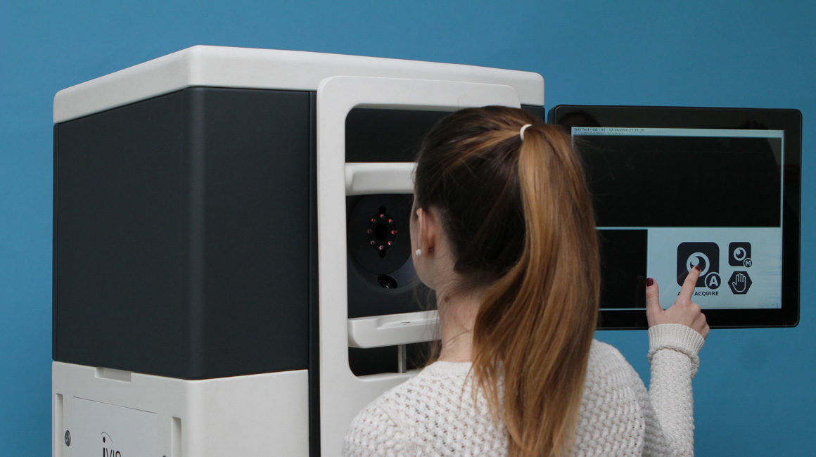
Fully automated acquisition, touch screen driven
Voice guided patient auto-positioning 3D eye-tracker synchronized with data system detection
Blue scanning laser micro-slit
Epithelial spatially registered maps
Ergonomic design
Intuitive software interface
Big data web based statistical analysis for custom vision surgery
Synchronous stereo high-resolution CMOS tandem cameras granting:
– Acquisition of over 120.000 data points per each detected surface
– High speed fully automated acquisition of 60 images in one second
– 3D synchronous tracking of all six spatial degrees of freedom
– Certified repeatability of the acquired maps
– Ocular features mapping for 3D intra-operative registration
– Ultrathin blue laser slit for high resolution images
– Optical stop-passing filters to remove parasite lights
– Automated voice guided system of patient positioning and acquisition 3D motorized chinrest
– Touch screen monitor and intuitive user interface
– Realtime exams backup
– Custom corneal refractive and therapeutic surgery
– Morphological and refractive analysis of the anterior segment of the eye
– Diagnostic analysis and clinical follow up of corneal pathologies
– Surgical outcomes and surgical follow up for corneal refractive and therapeutic procedures
Exam procedure:
Full automated voice driven acquisition and validation
Acquired data: 60 frames in 1 second with over 120000 points detected per each detected surface
Accuracy: ≤ 3um in the central 6 mm (diameter)
Field of view: 10mm
Image acquisition system: 2 Gigabit CMOS digital cameras
Eye tracking: 3D synchronous tracking of all six spatial degree of freedom
Slit light source: Ultra-thin blue laser diode
Laser diode slit divergence: ≤ 10% within 3mm depth of field
Weight: 135 kg
Power requirements: 110 ÷ 240Vac, 50-60Hz, 0.9A
Dimensions: 590mm x 485mm x 1565mm (LxWxH)
– Elevation maps: anterior, stromal and posterior
– Thickness map: epithelium, stromal and total
– Anterior chamber depth and iris maps
– Ray tracing refractive map: anterior, stromal, posterior and total
– Axial refractive map: anterior, stromal, posterior and total
– Tangential refractive map: anterior, stromal, posterior and total
– Mean power refractive maps: anterior, stromal, posterior and total
– Refractive aconic fitting: anterior, stromal, posterior and total
– Corneal morphological repeatability analysis: anterior, posterior and total
– Corneal refractive repeatability analysis: anterior, posterior and total
– 6 degrees of freedom eye-tracker display
– Horizontal and vertical angle K
– Fixation score
– Measuring tool
– Medical record
– Custom maps display
– Relevant data point display
– Diagnostic analysis and clinical follow-up
– Surgical outcomes and surgical follow-up


